800+
Brain regions

The complete end-to-end platform for quantitative 3D mouse brain imaging, combining whole brain immunolabelling, light sheet microscopy, and AI-assisted image analysis. Use every module or only the steps you need.
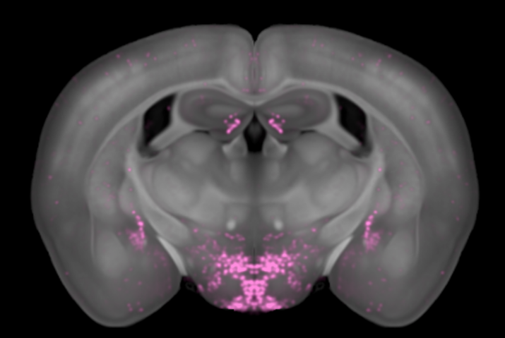
From tissue clearing and light sheet microscopy to spatial transcriptomics, single-cell sequencing and traditional immunohistochemistry, neuroscientists today have more tools than ever to study the brain. The challenge is no longer access to technology. It's knowing which approach will actually answer your research question. Each method has trade-offs in resolution, throughput, coverage and cost, and choosing the wrong one can mean months of work that misses the biology that matters most. Whole-brain imaging is transforming how researchers map activity, track drug distribution and uncover circuitry, but it's not the right fit for every question. Take the test to find out whether whole-brain imaging can accelerate your research.
A modular preclinical imaging platform that transforms a mouse brain sample into a complete, quantitative 3D dataset covering every anatomical region simultaneously, with AI-automated cell detection, atlas registration, and interactive delivery.
Brain regions
Data per brain
AI-assisted analysis
Interactive data
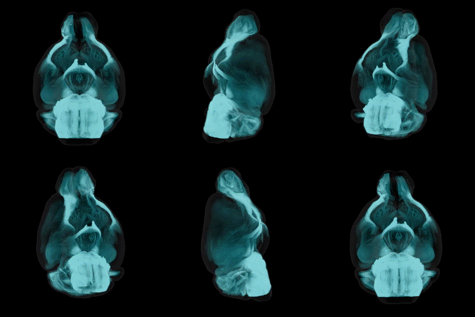
Each module can be used independently or combined. The platform is designed so that the output of each step feeds directly into the next, but you can enter or exit at any point.
Module 01
3D IHC staining of intact mouse brains and optical tissue clearing using iDISCO. Compatible markers include c-Fos, CD31, aSMA, pSTAT3, GFP, RFP and custom antibodies.
Standalone use: receive cleared, stained brains returned to your lab.
Module 02
Full 3D acquisition of cleared brains at cellular resolution using light sheet fluorescence microscopy. Generates ~1 TB of raw image data per brain sample.
Standalone use: imaging of externally labelled and cleared samples.
Module 03
Deep learning cell detection, signal quantification, and CCF atlas registration. Delivering quantitative results for 800+ brain regions via CNS-Voyager™.
Standalone use: analysis of your raw data and upload to CNS-Voyager™.
Add on
Scientific study reports and IND-enabling regulatory reports available on request for any module combination. Suitable for FDA/EMA submissions.
Available on request: on any study, any module combination.
VIBRAINT PRODUCT SERIES - EPISODE 1
Hosted by Harry Salt (neuroscientist & life sciences content creator) · Guest: Jacob Hecksher-Sørensen, CEO & Co-founder, Vibraint · Topics: what LS-Journey™ is · the modular service model · how data is delivered · open science with CNS-Voyager™ · global logistics · the virtual brain vision
Available on Spotify & YouTube

"Because this data is so rich. There's so much data. You can almost only view it in an online platform. Here you can scroll through the brain in any dimension you want and look up any brain region. You have the whole data at your hands."
-Jacob Hecksher-Sørensen, Vibraint
"One of the main goals for whole brain imaging is that you want to have an unbiased view of what happens. Maybe the most interesting effects you see are actually not where you initially thought you would see something."
-Jacob Hecksher-Sørensen, Vibraint
"Our hope is that once you have finalised the study and published the data, you make your maps available in the open access part of the viewer, so everybody can have a look and get inspired from those data sets."
-Jacob Hecksher-Sørensen, Vibraint
Whole brain immunolabelling combines optical tissue clearing with three-dimensional immunohistochemistry (IHC) to label specific proteins throughout an entire intact mouse brain. Rather than cutting the brain into 20–40 µm sections and examining a handful of pre-selected regions, this technique captures every labelled cell across all 800+ anatomical regions simultaneously, all in a single, unbiased 3D experiment.
The brain must first be processed to remove lipids. Primary antibodies then diffuse through the full tissue volume over 7–8 days at 37°C before secondary fluorescent antibodies complete the labelling. The result is a cleared, fluorescently-labelled brain ready for light sheet imaging.
10–15 regions sampled. Pre-selected. Sampling bias unavoidable
Brain destroyed. Sections cut. 3D spatial context permanently lost.
Manual counting. Weeks of work per brain. Observer-dependent.
800+ regions simultaneously. All brain regions captured at once.
Intact 3D volume . Full spatial context. Anatomy preserved in 3D
AI assisted quantification. Every cell detected automatically. Reproducible
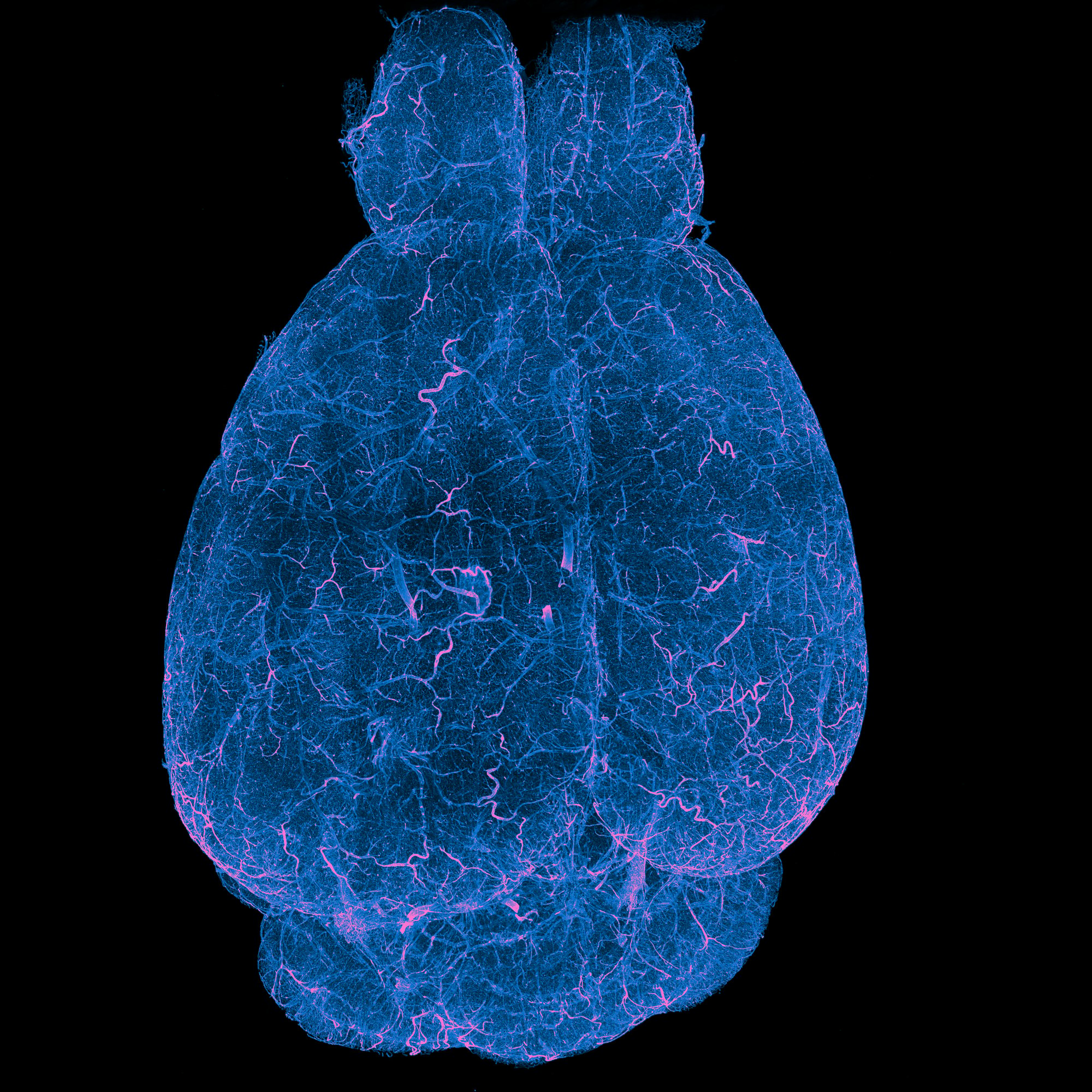
Methanol dehydration → DCM delipidation → DBE refractive-index matching. Fast, high transparency. Gold standard for whole brain antibody-based IHC.
Click here for more information about tissue processing and access to Vibraint's iDISCO protocol
iDISCO+ is compatible with a broad range of fluorescent markers used in preclinical neuroscience. Immediate early genes (IEGs) such as c-Fos, Arc, Egr1, and FosB are among the most widely used, reporting neuronal activity following pharmacological or behavioural stimuli. Cell-type markers enable brain-wide quantification of specific populations: NeuN for mature neurons, TH for dopaminergic neurons, ChAT for cholinergic neurons, Iba1 for microglia, and parvalbumin or calbindin for inhibitory interneuron subtypes. Signalling and phosphorylation markers, including pSTAT3, pERK, and pCREB, and receptor activation pathways, are particularly valuable for drug mechanism-of-action studies. Structural markers such as MBP and Neurofilament allow whole-brain mapping of myelin and axonal architecture, while vascular markers including CD31 enable cerebrovascular mapping. For disease pathology, beta-amyloid, phospho-tau, and alpha-synuclein have been applied in Alzheimer's and Parkinson's models. For transgenic reporter lines expressing GFP, RFP, or their derivatives, native fluorescence is partially quenched during iDISCO+ processing, but the signal can be reliably recovered by immunostaining with anti-GFP or anti-RFP antibodies, preserving the full benefits of whole-brain 3D imaging. The same pipeline also maps drug biodistribution and blood-brain barrier passage using fluorescently labelled compounds.
Neuronal activity/inhibition mapping
c-Fos, pPDH, Arc, Egr1, FosB
Markers against specific cell types
TH, Iba1, NeuN, ChAT
Receptor activation pathways
pSTAT3, pERK, pCREB
Connectivity and genetic reporters
GFP, RFP, tdTomato, mCherry
Architecture mapping
CD31, aSMA, MBP, NF
Markers of disease pathology
aBeta, aSyn, pTau
At Vibraint, we are always looking to push the boundaries of whole brain imaging. If you have a custom marker you would like to explore, we welcome the opportunity to collaborate.
Light sheet fluorescence microscopy (LSFM) illuminates the cleared brain with a thin laser sheet. One plane at a time. A camera positioned perpendicular to the light sheet captures the fluorescent signal from that plane, and the brain is scanned through the full volume to build a complete 3D image stack at single cell resolution.
Unlike confocal microscopy, which scans point-by-point, LSFM illuminates and captures entire planes simultaneously, making it orders of magnitude faster and causing minimal photobleaching. A full mouse brain can be imaged in hours rather than days.
The result is approximately 1 terabyte of raw image data per brain. A dataset so large that most research institutions lack the storage, computing infrastructure, and analysis pipelines to process it independently. This is the gap that LS-Journey™ Modules 02 and 03 address together.
Module 02 standalone service
Vibraint can image externally cleared brain samples. You can also submit your own raw light sheet data for AI analysis in Module 03, without engaging Module 02.
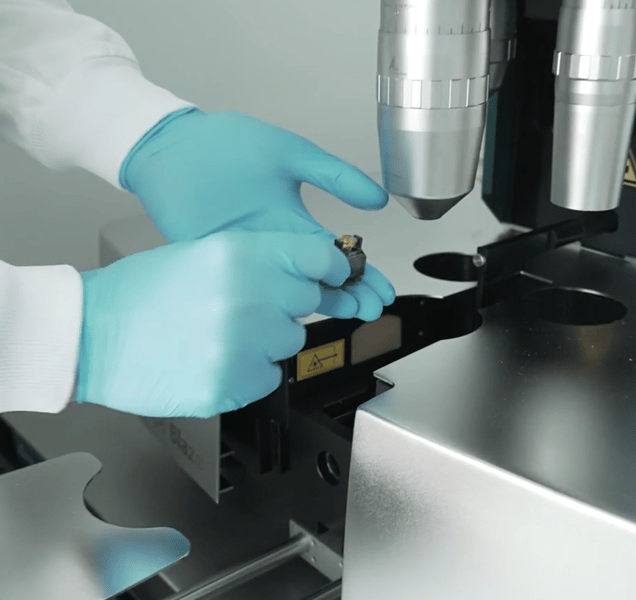
Setting up the microscope
The optically transparent brain from Module 01 is mounted in an imaging chamber filled with a refractive-index matching medium (ECi or equivalent) that matches the microscope objective.
Plane-by-plane laser sheet illumination
A thin sheet of laser light (typically 3–5 µm) illuminates one optical plane of the brain at a time. The illumination sheet is perpendicular to the detection axis. Only the focal plane fluoresces, minimising out-of-focus background.
Wide-field camera captures the plane
A wide-field sCMOS camera captures the full illuminated plane in a single frame or in multiple tiles, making acquisition orders of magnitude faster than point-scanning confocal methods.
Z-stack builds complete 3D volume
The brain is scanned through its full depth (~10 mm), building a complete 3D image stack at cellular resolution. Raw output: ~1 TB of image data, passed to Module 03 for AI analysis.
One terabyte of raw light sheet images contains millions of individually labelled cells distributed across hundreds of brain regions. Extracting quantitative, reproducible numbers from this data by hand is practically impossible. LS-Journey™ Module 03 uses deep learning AI pipelines to automate every step of the analysis, producing publication-ready quantitative results.
Every dataset is registered to Vibraint's published mouse brain Common Coordinate Framework (CCF), a peer-reviewed 3D reference brain atlas that enables consistent anatomical mapping across samples, cohorts, and imaging modalities. The CCF was developed and published in Neuroinformatics by Vibraint founders Johanna Perens and Jacob Hecksher-Sørensen.
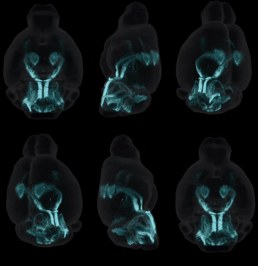
Results are delivered online via CNS-Voyager™, Vibraint's interactive 3D brain platform. Here researchers can explore their data, overlay public datasets (connectivity, transcriptomics, activity), and export figures for publication.
Module 03 standalone service
Submit your externally acquired light sheet dataset (from any instrument) for AI analysis and CCF registration. Results delivered via CNS-Voyager™.
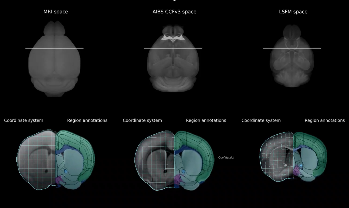
Perens et al., 2021. An Optimized Mouse Brain Atlas for Automated Mapping and Quantification of Neuronal Activity Using iDISCO+ and Light Sheet Fluorescence Microscopy. Neuroinformatics
.
Perens et al., 2023. Multimodal 3D Mouse Brain Atlas Framework with the Skull-Derived Coordinate System. Neuroinformatics 21(2):269–286.
VIBRAINT PRODUCT SERIES - EPISODE 3
Hosted by Harry Salt (neuroscientist & life sciences content creator) · Guest: Johanna Perens, Co-founder & CTO, Vibraint
Topics: the analysis bottleneck · why brain image analysis is an interdisciplinary challenge · deep learning and classical segmentation · model fine-tuning per experiment · region-wise and voxel-wise analysis · choosing the right endpoint · CNS-Voyager™ as an exploration tool
60 min · Available on Spotify & YouTube

"I do not believe in high-throughput pipelines that do not do any QC. If you do not check what comes out, you do not actually know if you found all the cells."
-Johanna Perens, Vibraint
"Every time you get a new experiment, you need to evaluate how similar it is to data before. There are no foundational AI models for light sheet data. You need to fine-tune the model, and you need to quality-check, every single time."
-Johanna Perens, Vibraint
"We see CNS-Voyager as a guiding tool for the whole experimental process. You can use public datasets to generate hypotheses, perform a study, find interesting areas, and do follow-up experiments. It becomes part of the neuroscientist's toolbox."
-Johanna Perens, Vibraint
Deep learning cell detection
A trained deep learning model scans the 3D volume and detects individual fluorescently-labelled cells. Output: 3D coordinates and signal intensity for every detected cell across the full brain volume.
CCF atlas registration
The light sheet dataset is non-linearly registered to Vibraint's published 3D mouse brain CCF. This maps every detected cell to its correct anatomical region enabling region-specific quantification and cross-sample comparison.
Region-specific quantification
Cell counts and signal intensity are aggregated per anatomical region, producing quantitative results for all 800+ regions simultaneously. Outputs include cell density maps, activity heatmaps, and region-level statistical tables.
CNS-Voyager™ interactive delivery
All results delivered online via CNS-Voyager™, allowing 3D and 2D interactive brain exploration, stereotactic coordinate placement, public dataset overlays, and export of publication-ready figures. No software installation required.
Vibraint's segmentation pipeline combines classical image processing with deep learning, using each where it performs best. Classical algorithms generate initial segmentations, which are manually curated by our scientists to create high-quality training data. Deep learning models are then trained or fine-tuned on this curated data, learning the signal in its full context: regional variation, tissue artifacts, and cross-instrument variability.
For every new experiment, we evaluate how similar the data is to our accumulated reference database and determine whether an existing model generalises or requires adaptation. There are no foundational AI models for light sheet data, and we do not apply models without verifying their performance on your specific dataset.
Quality control is performed at every processing step: data curation, segmentation, registration, and quantification. We check what comes out of every pipeline stage before it reaches your results.
If your lab or core facility already generates light sheet data, you do not need to use the full LS-Journey™ pipeline. Module 03 is available as a fully standalone service: you send us your raw light sheet images, and we return quantitative, atlas-registered results delivered interactively via CNS-Voyager™.
This is the fastest route from existing data to publishable endpoints. Our analysis pipeline handles data from any light sheet microscope architecture. We accept TIFF and HDF5 formats, set up a secure cloud bucket for your data transfer, and work with you to define the right segmentation approach and quantitative endpoints for your specific markers and biological question.
Every dataset is registered to the peer-reviewed Perens CCF, producing quantitative results across 800+ brain regions. Deep learning models are fine-tuned for your data, with quality control at every step. You receive cell counts, densities, signal coverage, intensity measurements, and voxel-wise statistical maps, all explorable in 3D through CNS-Voyager™.
For many research groups, this is the most practical way to access whole-brain quantitative analysis without building an in-house pipeline. You keep control of your tissue processing and imaging, and we handle the part that requires specialized computational neuroscience expertise.
The imaging settings you choose determine the endpoints you can measure. Resolution, step size in Z, and channel selection all constrain downstream analysis. A decision that seems reasonable at the microscope can make certain quantitative endpoints impossible to extract.
Our scientists consult before your study begins, ensuring your scan parameters, marker selection, and tissue processing are optimized for the analysis you need. This applies whether you use Vibraint for the full LS-Journey™ pipeline or only for analysis of your own externally acquired data.
Scientific study reports and regulatory-formatted IND-enabling reports are available as add-ons for any LS-Journey™ study, across any combination of modules.
A complete scientific summary of your LS-Journey™ study, presenting the quantitative brain imaging data in a structured, publication-ready format authored by Vibraint's team of neuroscience imaging scientists.
Narrative summary of key findings across brain regions
Quantitative data tables. Cell counts per region, statistical analysis
High-resolution figures and whole-brain activity maps
Interpretation relative to the CCF anatomical framework
Methods section describing the LS-Journey™ pipeline
A regulatory-formatted report for whole brain imaging data. Structured for inclusion in Investigational New Drug (IND) applications to the FDA, EMA, or equivalent regulatory authorities. Suitable for CNS drug development programs requiring preclinical brain imaging data.
Systematically formatted per regulatory documentation standards
Complete methodology documentation
Structured quantitative brain imaging data, target engagement, off-target activity
Documentation of QC steps
Statistical analysis and between-group comparisons
LS-Journey™ runs on methods that Vibraint's founders developed and published while working in CNS drug discovery at Novo Nordisk and Gubra, before founding Vibraint. The six papers below cover the whole pipeline: where a drug goes in the brain, which neurons respond, and how every result is registered to a light sheet reference atlas. The founders' whole-brain GLP-1 distribution work at Novo Nordisk also supported the NDA for Saxenda (liraglutide), so whole-brain imaging data has already carried weight in a regulatory submission.
The arcuate nucleus mediates GLP-1 receptor agonist liraglutide-dependent weight loss
Secher A, Jelsing J, Baquero AF, Hecksher-Sørensen J, ..., Knudsen LB. J Clin Invest 2014;124(10):4473–4488.
Fluorescent liraglutide and light sheet imaging showed that a peripherally dosed GLP-1 peptide reaches the arcuate nucleus behind the blood-brain barrier, in a receptor-dependent way. The same approach runs in LS-Journey™ biodistribution studies.
Integrated brain atlas for unbiased mapping of nervous system effects following liraglutide treatment
Salinas CBG, Lu TT, Gabery S, Marstal K, Alanentalo T, Mercer AJ, Cornea A, Conradsen K, Hecksher-Sørensen J, Dahl AB, Knudsen LB, Secher A. Sci Rep 2018;8:10310.
Drug distribution, whole-brain c-Fos, receptor expression and 2,469 Allen connectivity maps were combined in one atlas, separating direct drug effects from secondary activation. This is where integrative spatial biology started.
Whole-brain activation signatures of weight-lowering drugs
Hansen HH, Perens J, Roostalu U, ..., Hecksher-Sørensen J. Mol Metab 2021;47:101171.
Whole-brain c-Fos signatures of six weight-lowering drug classes, including semaglutide and setmelanotide, were mapped across 308 brain regions. All six activated overlapping feeding centers in the dorsal vagal complex and hypothalamus, plus limbic and dopaminergic regions. The same whole-brain comparison is available in LS-Journey™ c-Fos studies.
An optimized mouse brain atlas for automated mapping and quantification of neuronal activity using iDISCO+ and light sheet fluorescence microscopy
Perens J, Salinas CG, Skytte JL, ..., Hecksher-Sørensen J. Neuroinformatics 2021;19:433–446.
Introduced the Perens CCF, a reference atlas built from 139 iDISCO+ cleared brains that maps c-Fos faster and more accurately than the Allen CCFv3, especially in the hindbrain. Every LS-Journey™ dataset is registered to it, and the atlas now covers 800+ annotated regions.
Multimodal 3D mouse brain atlas framework with the skull-derived coordinate system
Perens J, Salinas CG, Roostalu U, Skytte JL, Gundlach C, Hecksher-Sørensen J, Dahl AB, Dyrby TB. Neuroinformatics 2023;21:269–286.
Linked MRI, Allen CCFv3 and light sheet templates with bidirectional deformation fields, plus a skull-derived stereotaxic coordinate system. LS-Journey™ results can be compared with public datasets and targeted again in living animals.
Digital brain maps and virtual neuroscience: an emerging role for light-sheet fluorescence microscopy in drug development
Perens J, Hecksher-Sørensen J. Front Neurosci 2022;16:866884.
Set out how compact, atlas-aligned digital brain maps can be shared and compared in the browser to speed up CNS drug development. It is the model for delivering LS-Journey™ results in CNS-Voyager™.
A full LS-Journey™ study delivers every output below. Individual modules deliver their respective subset. You choose the combination.
Anatomical segmentation
All 800+ mouse brain regions identified and labelled per the CCF. A reproducible structural framework across your cohort.
Quantitative cell counts
Region-specific counts for all labelled cell populations and signal intensities. Exportable tables, heatmaps, and statistical-analysis-ready data files.
Whole-brain activity maps
Immediate early genes (IEGs). Quantified across the full brain, CCF-registered for direct cross-sample and cross-cohort comparison.
CNS-Voyager™ 3D viewer
Interactive online brain exploration. 3D and 2D navigation, stereotactic coordinates, region lookup, figure export.
Multi-modal integration
Overlay your data with Allen Brain connectivity, spatial transcriptomics, and public activity datasets. All in the same reference space.
Publication-ready outputs
High-resolution brain figures, statistical summaries, and data exports formatted for peer-reviewed neuroscience journals.
Study report (on request)
Scientific narrative summary of findings, quantitative tables, figures, and statistical interpretation. Authored by Vibraint scientists.
IND report (on request)
Regulatory-formatted report for FDA/EMA IND submissions.
Comprehensive answers covering all modules
LS-Journey™ is Vibraint's modular whole brain imaging platform for mouse models, combining whole brain immunolabelling (optical clearing and 3D IHC staining), light sheet fluorescence microscopy, and AI-assisted image analysis with CCF atlas registration. Results are delivered interactively via CNS-Voyager™. Each module can be used independently or combined. Study reports and IND-enabling regulatory reports are available as add-ons for any study.
Whole brain immunolabelling combines optical tissue clearing with 3D IHC to label proteins across an intact mouse brain, capturing all 800+ regions in a single unbiased experiment. Traditional IHC sections the brain and samples 10–15 pre-selected regions, introducing sampling bias, destroying 3D spatial context, and requiring weeks of manual counting. Whole brain immunolabelling eliminates all three limitations.
Light sheet microscopy illuminates one plane of a cleared brain at a time with a thin laser sheet. A perpendicular camera captures each plane as the brain is scanned through its full depth, building a 3D volume at cellular resolution. This generates ~1 TB of raw data per brain with minimal photobleaching. Acquisition takes hours rather than days, making whole-brain cellular-resolution imaging practical for large studies.
Deep learning algorithms automatically detect and count every labelled cell in the 3D light sheet volume. The dataset is then registered to Vibraint's published mouse brain CCF, mapping each detected cell to its anatomical region. Output includes quantitative cell counts for 800+ regions, whole-brain activity maps, and anatomical segmentation. All delivered via CNS-Voyager™.
Vibraint uses a light sheet Common Coordinate Framework (CCF) developed by founders Johanna Perens and Jacob Hecksher-Sørensen and published in Neuroinformatics (Perens et al., 2021). This peer-reviewed 3D atlas enables reproducible anatomical registration across all samples, cohorts, and imaging modalities.
Yes. LS-Journey™ is fully modular. You can use the complete pipeline or individual modules independently. For example: Vibraint can perform AI analysis on your own externally acquired light sheet data (Module 03 only); or clear and stain your brains and return them to your lab for imaging (Module 01 only). Study reports and IND-enabling reports can be added to any combination.
Compatible markers include immediate early genes (c-Fos, pPDH), cell-type markers (Iba1, TH, ChAT, Parvalbumin), signalling markers (pSTAT3, pERK), transgenic reporters (GFP, tdTomato, mCherry), and structural markers (CD31, MBP, NF). Custom antibody targets can be validated on request. Contact Vibraint at the quote stage.
We accept TIFF and HDF5 (.h5) files, the standard outputs of most light sheet microscopes. Data is shared via a secure cloud bucket that we create for each customer, with upload guidelines provided. If you are unsure whether your data format is compatible, contact us at the quote stage.
We recommend a minimum of eight animals per group for robust statistical analysis. Studies with six animals per group are possible but come with reduced statistical power, which we note transparently in the results. The optimal sample size depends on your expected effect size and biological question. We can discuss this during study design consultation.
The Perens CCF is built from adult male C57BL/6 mice processed with iDISCO+. Registration is most reliable when samples closely match this reference. For different strains, sexes, ages, or tissue processing protocols, we apply automated registration quality control and, where needed, generate protocol-specific brain templates linked to the CCF via validated transformation chains. We recommend discussing your specific model during study design.
Yes. We offer consultation sessions for researchers who want to set up or improve their own analysis workflows using open-source tools such as BrainGlobe. We can advise on pipeline architecture, parameter selection, quality control strategies, and endpoint design, drawing on our experience processing hundreds of whole-brain datasets across diverse markers and protocols.
Tell us your target markers, mouse model, sample numbers, and reporting needs and our scientists will design the optimal module combination and provide a detailed quote.