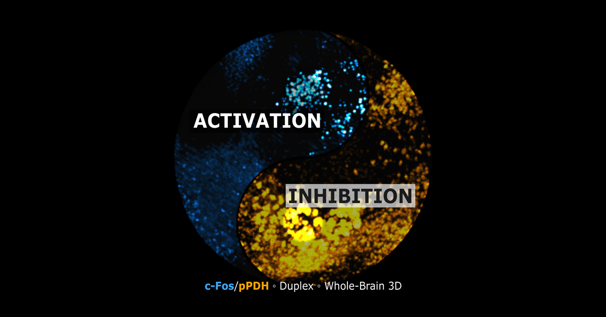
pPDH is pyruvate dehydrogenase E1 alpha phosphorylated at Ser293, the inactive form of the enzyme that feeds pyruvate into the TCA cycle. Yang et al. (Neuron, 2024) showed that pPDH rises when neuronal firing decreases and falls when it increases, making it the first endogenous marker that can be stained in tissue to show where neurons went quiet, the inverse of what c-Fos shows for where neurons became active.
Why neuroscience needed a marker like this
c-Fos and the other immediate early genes have given whole-brain activity mapping a reliable readout of activation for decades, but they are silent on inhibition. A neuron that stops firing does not turn on an immediate early gene. That leaves half of any circuit's story untold: a brain region could look unremarkable on a c-Fos map simply because its activity decreased rather than because nothing happened there at all. pPDH closes part of that gap by giving inhibition its own measurable signal, read directly from brain tissue rather than inferred from the absence of activation.
How pPDH was validated
Yang et al. 2024 showed pPDH rising in mice in several independent situations where neuronal activity was known to drop: after general anesthesia, after chemogenetic silencing of specific neurons, at the end of a period of visual stimulation, after drinking resolved a period of thirst, and after refeeding ended a period of fasting. In each case, the brain regions expected to go quiet showed a pPDH increase, and the signal receded once activity returned to baseline.
How quickly pPDH responds, compared with c-Fos
pPDH moves faster than c-Fos in both directions. In mice, pPDH was clearly elevated within 1 hour of anesthesia onset or immediately after drinking ended a period of thirst, while c-Fos in the same thirst model took up to 8 hours to return to its own baseline. In cultured neurons, pPDH dropped within 10 minutes of stimulation and recovered roughly 50 minutes after the stimulation stopped. c-Fos, by contrast, typically takes longer to turn on and longer still to clear, which is why it captures a wider window of past activity while pPDH captures something closer to a brain's state at the moment of fixation.
Measuring c-Fos and pPDH in the same brain
The two markers do not compete for the same signal. c-Fos accumulates in the nucleus; pPDH is read in the cytoplasm. That physical separation, combined with reading each antibody in its own fluorescence channel, means both can be labeled and imaged in a single cleared mouse brain. The result is a brain-wide, two-channel map that shows where a circuit turned on and where it turned off in the same animal, rather than having to infer the inhibited side from a separate cohort or a different study.
Where pPDH does not work well
pPDH has known blind spots, and an honest marker is one that states them. In regions outside the blood-brain barrier, such as the area postrema and the subfornical organ, baseline pPDH is already high and does not respond to anesthesia. CA3 of the hippocampus and POMC neurons also showed no pPDH change in the validating study. Compounds that directly affect neuronal glucose metabolism need extra controls, since pPDH sits on a metabolic enzyme and a drug that shifts glucose handling independently of neuronal firing could confound the readout.
What is established, and what is still open
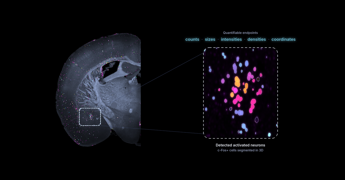
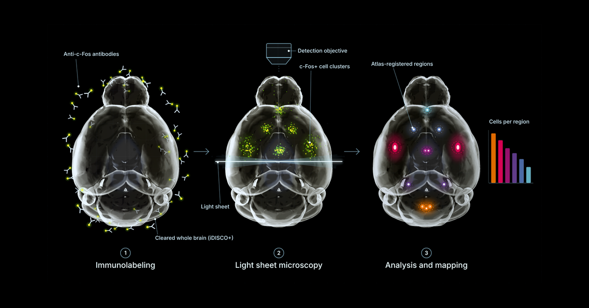
Whole-brain pPDH staining and mapping in the intact, cleared mouse brain is technically established: the antibody labels the protein, iDISCO+ clearing and light sheet microscopy image it brain-wide, and registration to a reference atlas maps it to specific regions the same way c-Fos is mapped today. What remains genuinely open is the biological interpretation of a given pPDH signal in a new experimental context, since the marker is only two years old in the literature and its behavior has been characterized in a specific set of paradigms so far, not exhaustively across every brain state and compound class. That is also why Vibraint is offering a collaboration discount on combined c-Fos and pPDH studies through the end of 2026: the fastest way to build a reliable picture of what pPDH does and does not capture is to run it across more studies and compare notes, in collaboration with the labs doing the work.
Frequently asked questions
What is pPDH?
pPDH is pyruvate dehydrogenase E1 alpha phosphorylated at Ser293, the inactive form of the enzyme that feeds pyruvate into the TCA cycle. Yang et al. (Neuron, 2024) showed that pPDH rises when neuronal firing decreases and falls when it increases, making it the first endogenous inverse activity marker that can be stained in tissue.
Is there a marker for neuronal inhibition, like c-Fos is for activation?
pPDH is the best-characterized candidate so far. It increased in vivo after general anesthesia, chemogenetic silencing, the end of visual stimulation, drinking after thirst and refeeding after fasting. It is still exploratory, with known blind spots in some regions and cell types.
Can c-Fos and pPDH be measured in the same brain?
Yes. The two antibodies are read in separate fluorescence channels, and the signals sit in different compartments: c-Fos in the nucleus, pPDH in the cytoplasm. Both can be labeled in one cleared mouse brain and mapped to the same reference atlas.
How quickly does pPDH respond compared with c-Fos?
Faster. In mice, pPDH was clearly elevated 1 hour after anesthesia or after drinking ended a period of thirst, while c-Fos took up to 8 hours to return to baseline in the same thirst model. In cultured neurons, pPDH dropped within 10 minutes of stimulation and recovered about 50 minutes after it stopped.
Where does pPDH not work well?
In regions outside the blood-brain barrier, such as the area postrema and subfornical organ, baseline pPDH is high and did not respond to anesthesia in the validating study. CA3 of the hippocampus and POMC neurons also showed no pPDH change. Compounds that directly affect neuronal glucose metabolism need extra controls.
Is whole-brain pPDH imaging available as a service?
Yes. Vibraint maps pPDH alongside c-Fos across the whole mouse brain through LS-Journey™, delivered interactively in CNS-Voyager™. A collaboration discount is available on combined studies submitted through the end of 2026.
Reference
Yang, Z. et al. Phosphorylation of pyruvate dehydrogenase inversely associates with neuronal activity. Neuron 112, 959-971 (2024). doi:10.1016/j.neuron.2023.12.015