logo_text_black_bg

MAP EVERY AXONAL PROJECTION ACROSS THE ENTIRE BRAIN

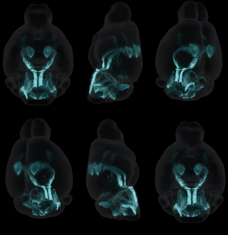
Brain-wide axonal tracing with light sheet fluorescence microscopy and AI-assisted CCF atlas registration. Map anterograde, retrograde, and trans-synaptic projections across all 800+ brain regions. All in a single 3D experiment.

iDISCO+ clearing · light sheet fluorescence microscopy · AI-assisted image analysis · CCF brain atlas registration · CNS-Voyager™ integration

section_5
TAKE THE TEST

FIND OUT IF WHOLE BRAIN IMAGING CAN ACCELERATE YOUR RESEARCH.

From tissue clearing and light sheet microscopy to spatial transcriptomics, single-cell sequencing and traditional immunohistochemistry, neuroscientists today have more tools than ever to study the brain. The challenge is no longer access to technology. It's knowing which approach will actually answer your research question. Each method has trade-offs in resolution, throughput, coverage and cost, and choosing the wrong one can mean months of work that misses the biology that matters most. Whole brain imaging is transforming how researchers map activity, track drug distribution and uncover circuitry, but it's not the right fit for every question. Take the test to find out whether whole brain imaging can accelerate your research.


TRACER TYPES

THREE CLASSES OF TRACER,
THREE DIFFERENT QUESTIONS

The choice of tracer determines what the experiment reveals. LS-Journey™ is compatible with all three classes, and Vibraint can advise on the optimal tracer for your research question.


Anterograde tracers

Where do my neurons send their outputs?

Taken up by neuronal cell bodies at the injection site and transported forward along axons to axonal terminals to reveal the complete output connectivity of the injected region.

AAV-XFP, BDA, PHAL

"Which brain regions receive input from my injection site?"

Retrograde tracers

Which neurons project to my region of interest?

Taken up at axonal terminals and transported backward to the cell body. Labelling all neurons that project to the injection site, regardless of their location in the brain.

retroAAV-GFP, CTB, FluoroGold, WGA

"Which regions send axons to my injection site?"

Trans-synaptic tracers

What is the multi-order circuit architecture?

Cross synaptic junctions to label neurons one or more synapses away from the injection site. Mapping the polysynaptic circuit structure of any brain region or cell population.

Modified rabies, HSV, PRV

"What is the full circuit architecture beyond my region?"

HOW IT WORKS

FROM TRACER INJECTION TO
BRAIN-WIDE MAP

LS-Journey™ is fully modular, so you enter or exit at any step. The most common entry point for connectivity studies is after the viral injection expression period.


TISSUE PREP

Perfusion, fixation and clearing

After the tracer expression period (typically 2–4 weeks for AAVs), the brain is perfusion-fixed. At Vibraint, the brains are processed for iDISCO+ using antibodies against the reporter protein (GFP, tdTomato).

Can accept pre-fixed brain tissue directly.

IMAGING

Light sheet fluorescence microscopy 

The cleared brain is scanned at 3–5 µm isotropic resolution. High enough to detect and quantify individual axonal fibres. Dual channel acquisition captures both the tracer signal and the autofluorescence channel for CCF registration.

Can accept external light sheet datasets.

AI ANALYSIS

Signal quantification & CCF registration

AI pipelines quantify axonal signal intensity and distribution across the 3D volume. The autofluorescence channel is registered to the Perens CCF, mapping every voxel of tracer signal to its correct anatomical region across all 800+ areas.

DATA DELIVERY

Interactive delivery on CNS-Voyager™

Projection maps, per-region signal density tables, and group-level comparisons delivered via CNS-Voyager™. Overlay with Allen Brain Atlas connectivity data and published activity maps for cross-modal insight.


ACADEMIC APPLICATION

WHAT WHOLE BRAIN CONNECTIVITY
MAPPING REVEALS

Because every brain region is captured in a single unbiased experiment, whole brain axonal tracing is transforming how academic neuroscientists study circuit architecture.

Connect_1-1

Input/output circuit mapping

Define the complete afferent and efferent connectivity of any brain region in a single experiment, without pre-selecting which structures to examine.


"What are the full inputs and outputs of the basolateral amygdala?"

Connect_2-1

Disease-related connectivity changes 

Compare axonal projection patterns between wild-type and disease model brains to identify structural connectivity changes associated with neurodegeneration or neuropsychiatric conditions.

"How does connectivity change in an Alzheimer's mouse model?"

Connect_3-1

Cell-type specific projection atlases

Using Cre-dependent AAVs, generate brain-wide projection maps restricted to a specific cell type. Dopaminergic, serotonergic, cholinergic, or any genetically defined population.


"Where do VTA dopamine neurons project across the brain?"

Connect_4-1

Multi-modal connectivity + activity integration

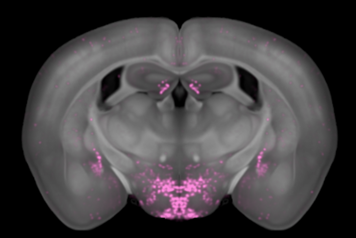
Overlay whole brain axonal tracing with c-Fos activity maps in the same CCF space, directly linking structural connectivity to functional engagement during a stimulus or treatment.

"Do regions with strong input also show elevated c-Fos after stimulation?"

Connect_5-1

Reproducible connectivity datasets for publication 

Generate quantitative, atlas-registered projection datasets that are directly comparable across animals, laboratories, and publications, supporting reproducible connectomics research.

"Can we produce a quantitative projection atlas of the lateral hypothalamus?"

DATA DELIVERY

YOUR CONNECTIVITY DATA. INTERACTIVE,
SHAReABLE, PUBLICATION READY

All LS-Journey™ connectivity data is delivered via CNS-Voyager™, Vibraint's interactive 3D brain platform. Explore your axonal projection maps in 3D and 2D, compare with the Allen Brain Atlas, and export figures for your next paper.

connectivity_web
  • 3D and 2D interactive axonal projection map viewer

  • Per-region signal density tables in 800+ anatomical regions

  • Overlay with Allen Brain Atlas connectivity reference data

  • Overlay with c-Fos activity maps in the same CCF space

  • Stereotactic coordinate lookup for injection site verification

  • Export high-resolution figures for publication

  • Shareable links for collaborators and reviewers
FREQUENTLY ASKED QUESTIONS

WHAT SCIENTISTS ASK ABOUT WHOLE
BRAIN CONNECTIVITY MAPS

What is whole brain axonal tracing?

Whole brain axonal tracing uses fluorescent tracer molecules injected into a specific brain region to label the axonal projections of neurons in that area. Combined with optical tissue clearing and light sheet microscopy, the complete 3D distribution of labelled axons can be mapped across all 800+ brain regions simultaneously. Revealing the full projection landscape of any brain area in a single unbiased experiment.

How does AAV tracing work with light sheet microscopy?

An AAV encoding a fluorescent protein (GFP, tdTomato, mCherry) is injected into the brain region of interest. After 2–4 weeks of expression, the protein fills infected neurons and their axons. The brain is perfusion-fixed, stained with antibodies against the reporter protein, and imaged with light sheet microscopy. AI analysis then registers the 3D dataset to the CCF and quantifies axonal signal intensity per anatomical region.

What is the difference between anterograde and retrograde tracing?

Anterograde tracers (AAV-GFP, BDA) are taken up by neuronal cell bodies and transported forward along axons to terminals. Revealing where injected neurons send their outputs. Retrograde tracers (CTB, FluoroGold, retroAAV) are taken up at terminals and transported back to the cell body, revealing which neurons project to the injection site. The choice depends on whether you want to map outputs (anterograde) or inputs (retrograde) of your region of interest.

What scanning resolution is needed for axonal tracing?

Axonal fibres typically require 3–5 µm isotropic resolution to reliably detect and quantify individual fibres and terminal fields. This is higher than the 5 µm sufficient for cell body detection (e.g. c-Fos). Vibraint optimises resolution settings for each connectivity study based on the tracer, fluorescent label, and analysis requirements.

Can I use Cre-dependent AAVs with LS-Journey™ connectivity mapping?

Yes. Cre-dependent (FLEX/DIO) AAVs that require Cre recombinase for expression are compatible with LS-Journey™ when used in Cre driver mouse lines. This enables cell-type specific projection mapping. For example, mapping the long-range projections of dopaminergic, cholinergic, or parvalbumin interneurons specifically. Contact Vibraint to discuss your Cre line and AAV serotype.

How is connectivity data registered to the mouse brain CCF?

The autofluorescence channel acquired during light sheet imaging serves as an anatomical fingerprint. This channel is non-linearly registered to Vibraint's published mouse brain CCF (Perens et al., Neuroinformatics 2021), placing the brain in a common coordinate space. All tracer signal is then mapped to this CCF, enabling region-specific quantification and cross-animal comparison.

Can I overlay whole brain connectivity data with c-Fos activity maps?

Yes. Because both connectivity data and c-Fos activity data from LS-Journey™ are registered to the same CCF, they can be directly co-visualised in CNS-Voyager™. This allows researchers to correlate structural connectivity with functional engagement. For example, asking whether regions that receive strong axonal input also show elevated neuronal activity after a stimulus or pharmacological intervention.

VIBRAINT'S LS-JOURNEY™ PLATFORM

LS-Journey™ is purposely designed to be modular, allowing you to customise your studies as needed. If you do the staining and microscopy, we can analyse the data and upload to CNS-Voyager™.

Tissue_processing
Microscopy
Analysis

READY TO DESIGN YOUR WHOLE BRAIN STUDY WITH LS-JOURNEY™

Tell us your target markers, mouse model, sample numbers, and reporting needs, and our scientists will design the optimal module combination and provide a detailed quote.