Skip to content
logo_text_black_bg
Whole brain c-Fos in response to Ozempic
c-Fos whole-brain imaging study design

HOW TO BEST DESIGN YOUR C-FOS STUDY FOR WHOLE BRAIN IMAGING

Jacob Hecksher-Sørensen
Johanna Perens
Thomas Topilko
Jacob Hecksher-Sørensen, Johanna Perens, and Thomas Topilko

Practical guidance from the scientists who have mapped thousands of brains. 

c-Fos whole-brain imaging has become one of the most powerful discovery tools in neuroscience. A single experiment can reveal which brain circuits respond to a drug, a behavioral challenge, or a disease state, across 800+ anatomical regions, in one pass. But the power of the readout depends entirely on how well the study is designed upfront.

Here is what we have learned from years of running whole-brain c-Fos studies, first at Novo Nordisk, Gubra, and Lundbeck, and now at Vibraint.

 

Understand the biology of your readout

"c-Fos is best seen as a brain-wide molecular snapshot. Extremely powerful for finding candidate circuits, but something that needs a clear follow-up with causal tools like optogenetics, chemogenetics, or electrophysiology."

-Thomas Topilko, Co-founder and CSO, Vibraint


When neurons become active, they switch on the immediate early gene c-fos, and the resulting protein accumulates in the nucleus like a flag indicating recent activity. The protein takes approximately 60 to 90 minutes to appear after neuronal activation and is then cleared over the following two to three hours.

Two practical consequences follow. First, the timing of animal sacrifice relative to your stimulus is the single most critical variable in your study design. Second, c-Fos is biased toward strong excitatory activity. Inhibitory neurons tend to express less c-Fos, and absence of signal does not prove absence of activity.


GET YOUR DOSING AND TIMING RIGHT

For pharmacological studies, the goal is to capture the peak of c-Fos induction after your compound reaches the brain. That requires accounting for two delays: the drug's own pharmacokinetics and the 60 to 90 minute c-Fos protein accumulation lag.

If you know your compound's tmax, dose at tmax and use a high dose to maximize signal. If your compound's PK is unknown, a practical default is a four-hour timepoint between dosing and perfusion.

Key numbers

60-90 min

c-Fos protein accumulation after neuronal activation

4 hours

Default dosing-to-perfusion timepoint when PK is unknown

CHOOSE THE RIGHT GROUP SIZE

Recommended group sizes

n=5-8

Pharmacological c-Fos studies (robust, reproducible responses)

n=8

Physiological c-Fos studies (behavioral, stress, sensory)

When in doubt, go higher rather than lower. The preferred way to address biological variability is to increase n, not to exclude animals after the fact.

"Studies with 5-6 animals per group are possible but come with statistical limitations."

-Johanna Perens, Co-founder and CTO, Vibraint


PERFUION QUALITY SETS THE CEILING

No amount of computational sophistication can rescue a badly perfused brain. The quality of your tissue at sacrifice determines everything downstream.

Key points from Vibraint's published tissue preparation protocol:

  • Use intracardiac perfusion with heparinized PBS followed by 4% PFA, both at room temperature.

  • Incise the right atrium first, then insert a blunt needle into the left ventricle.

  • Look for the fixation signs: tail curving and leg muscle stiffening. If absent, log a deviation.

  • Dissect the brain carefully. Avoid cuts, tears, and hair on the tissue.

  • Post-fix overnight in 4% PFA, then wash 3 x 30 min in PBS.

  • Store and ship at 4 degrees in PBS with 0.1% PFA. Never freeze.

Document everything at takedown. Take-down notes are the only basis on which an animal can be justifiably excluded later.


ANTIBODY CONSISTENCY IS NON-NEGOTIABLE

"The quality of the antibody you are using is absolutely critical. My rule is: once you find one that works, stick to it. Never change."

-Thomas Topilko, Co-founder and CSO, Vibraint

  • Use monoclonal antibodies. Lot-to-lot variation is negligible compared to polyclonal alternatives.

  • Consider directly conjugated primaries. Better signal-to-noise ratio by avoiding polyclonal secondary antibody background.

  • Stick with your antibody. Consistency across your dataset is worth more than marginal gains.

Process all brains from the same study together in one batch. When comparing results across separate studies, compare statistical maps that contrast treatment against control within each study, not raw cell counts directly.

This is also why a proper vehicle control group is essential in every study, not optional.


PLAN YOUR ANALYSIS ENDPOINTS BEFORE YOU START

"Before even starting the experiment, you need to know what you want to get out of it, to then know what settings to use for imaging. Every decision is a trade-off."

-Johanna Perens, Co-founder and CTO, Vibraint

Two complementary analysis types are typically run:

Region-based cell counts

c-Fos-positive cells counted per region, mapped to the Perens CCF, compared across 800+ regions

Voxel-wise statistical maps

Spatial "hotspots" of activity showing exactly where your drug has its strongest effect

THE DISCOVERY ADVANTAGE

With traditional histology, 10 to 20 µm slices represent roughly 0.1% of the brain. Whole-brain imaging flips this: you get the complete picture, and the biology you did not expect often turns out to be the biology that matters most.

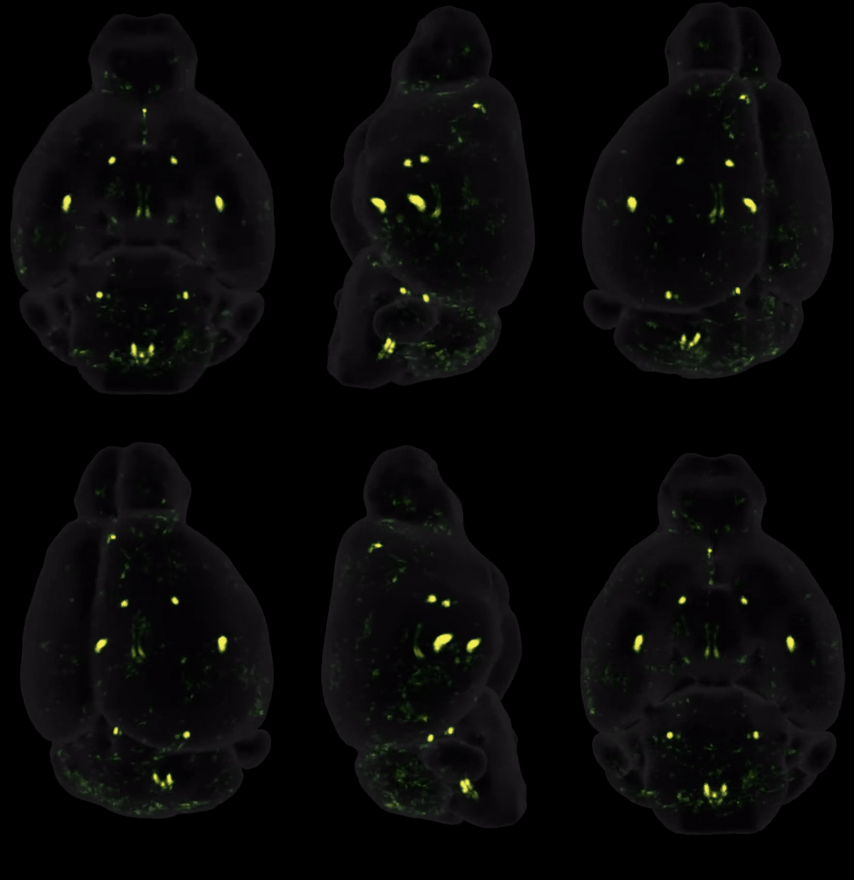
"What this map shows is not just a simple drug effect, but a window into how a metabolic signal, one that normally starts in the gut, becomes a brain-wide response."

-Thomas Topilko, Co-founder and CSO, Vibraint

RUN YOUR STUDY USING LS-JOURNEY™

With LS-Journey™, your fluorescence-intensity data is quantified across all 800+ brain regions, delivered as statistical maps with full group-level analysis, and made fully interactive in 3D and 2D on CNS-Voyager™. Studies can be acute or chronic, the latter revealing how distribution shifts as receptors downregulate over time. Because LS-Journey™ is modular, you can also send in externally generated light sheet data for AI-assisted analysis and upload to CNS-Voyager™.

START WITH A CONVERSATION

Planning a c-Fos study? Discuss your compound, research question, and timelines with our team. The right study design makes all the difference.