Skip to content
logo_text_black_bg

7 No-Code Ways to Make 3D Brain Poster Figures

Jacob Hecksher-Sørensen

 Here are seven no-code tools for making great 3D brain figures for your posters, papers, and grant applications, without writing a single line of code. 

1. Allen Reference Atlas 3D Viewer

Best for: Quick anatomical reference shots showing where a region sits in the brain.

The Allen Reference Atlas is the default starting point for most mouse brain work. The 3D viewer lets you rotate the brain, select individual regions from the CCFv3 annotation, and toggle transparency to show how deep structures relate to the surface. Works for adult mouse, developing mouse, and human brains.

Poster tip: Set the surrounding cortex to semi-transparent, highlight your region of interest in color, and screenshot. Clean, citable, and everyone on your poster committee will recognize the Allen atlas style.

Price: Free

2. Imaris / ZEISS arivis Pro

Best for: Cinema-quality 3D renderings of your own cleared-tissue or confocal data.

Imaris (Oxford Instruments) is the established standard for 3D visualization in neuroscience microscopy. Its GUI handles volume rendering, surface reconstruction, and filament tracing without any scripting. Load a light sheet or confocal Z-stack, and you can produce poster-ready 3D brain renders in minutes. ZEISS arivis Pro offers comparable capabilities and handles terabyte-scale datasets particularly well. Both require that you bring your own 3D data or a template brain to upload.

Poster tip: Use the Snapshot feature at 300 DPI with a black or white background for a clean poster panel. The Animation module can also produce rotating brain movies for digital posters or conference presentations.

Price: Commercial license (check your institution's imaging core for shared access)

3. BioRender

Best for: Clean, publication-ready schematic brain figures and graphical abstracts.

BioRender is not an atlas tool. It is an illustration platform with drag-and-drop neuroscience templates, including sagittal brain sections, circuit schematics, and cell-type icons. If you need a polished conceptual figure rather than data-driven 3D rendering, BioRender is the fastest path to something that looks professional.

Poster tip: Use BioRender for your mechanism or hypothesis panel, then pair it with a data-driven 3D figure from one of the other tools on this list. The combination of schematic and real atlas data tells a stronger story than either alone.

Price: Free tier available; full access through many institutional subscriptions

4. Scalable Brain Atlas

Best for: Comparing regions across species and atlases.

The Scalable Brain Atlas hosts over twenty publicly accessible brain atlases across mouse, rat, macaque, marmoset, human, ferret, and opossum. Its 3D mesh viewer lets you select and highlight regions, view them from any angle, and compare how the same structure is defined across different atlasing efforts.

Poster tip: If your poster bridges mouse and human data, or compares two different mouse atlases, this is the only free tool that lets you show both in the same visual framework.

Price: Free

5. Neuroglancer

Best for: Viewing and screenshotting high-resolution volumetric brain datasets.

Neuroglancer is a WebGL-based viewer developed at Google and the Seung Lab (Princeton). It handles massive volumetric datasets, including serial electron microscopy, light sheet volumes, and fMRI-scale data. Many public brain datasets (Allen, BRAIN Initiative, Brain Image Library) offer direct "Open in Neuroglancer" links.

Poster tip: Neuroglancer is powerful but takes some getting used to. The interface is not immediately intuitive. Budget 20 minutes the first time to learn the layer controls and camera. Once you have a good view, right-click to copy a shareable link so you can return to the exact same angle.

Price: Free

6. Fiji/ImageJ 3D Viewer

Best for: Making 3D renderings from your own confocal or light sheet Z-stacks.

If you have your own imaging data and want a 3D rendering for your poster, the 3D Viewer plugin in Fiji is the classic solution. Open your Z-stack, launch the 3D Viewer (Plugins > 3D Viewer), and it generates a volume rendering or surface model. You can rotate, crop, adjust colors, and export at any angle. No scripting required.

Poster tip: Import your cleared-tissue or confocal stack, set the rendering mode to "Volume," adjust the transfer function to bring out your signal, and export at 300 DPI. Fiji is already on most lab computers, so this takes minutes once you know the steps.

Price: Free, open source

7. CNS-Voyager™

Best for: Interactive 3D mouse brain figures with gene expression, connectivity, and stereotactic coordinates in one place.

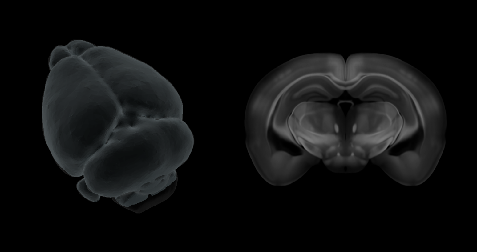
CNS-Voyager is a browser-based 3D mouse brain platform built on the peer-reviewed Perens CCF reference brain. Where the other tools on this list each do one thing well, CNS-Voyager integrates multiple data layers in a single viewer: 800+ anatomically defined brain regions, full Allen Brain Cell Atlas gene expression maps for every gene, published connectivity data, spatial transcriptomics overlays, and built-in stereotactic coordinate lookup.

This means you can build a poster figure that shows, for example, your target region highlighted in 3D with the expression pattern of your gene of interest overlaid, and the injection coordinates you used marked on the same brain. One screenshot, one tool.

Poster tip: Use the gene expression overlay to add biological context to your region of interest. A figure showing "this is where we injected, and here is where the receptor is expressed brain-wide" is far more compelling than a region highlight alone. You can also look up your stereotactic coordinates directly in the viewer, making it a daily-use tool beyond poster season.

Price: EUR 25/month for PhD students; EUR 100/month standard. Requires a WebGPU-capable browser.

comparison_table_mobile_1

Bottom line

You do not need to learn Python or spend a week troubleshooting a pipeline to put a credible 3D brain figure on your poster. For a quick anatomical reference, the Allen viewer is unbeatable. For cinema-quality renders of your own data, Imaris or arivis do the heavy lifting. For a polished schematic, BioRender does the job. For rendering your own stacks on a budget, Fiji is already on your computer.

If you want gene expression context, connectivity data, and stereotactic coordinates layered onto the same 3D brain, CNS-Voyager puts all of that in one browser tab, at a price designed for trainees.

Whichever tool you pick, your poster deserves better than a 2D atlas screenshot pasted from a PDF.