c-Fos
whole-brain imaging
LS-Journey™
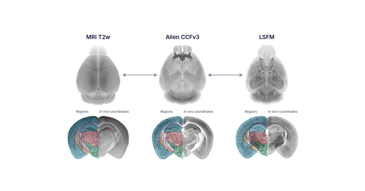

An atlas built for one imaging method does not automatically register well to another. Here is what actually changes when the method changes, and why it matters most where you are counting activated cells.
A common coordinate framework, or CCF, is a 3D reference brain in which every anatomical structure has already been defined and labeled. Registering a new brain image to that reference assigns each voxel to a named region, which turns a raw image into a set of region-level measurements you can compare across animals, studies, and labs.
Coverage varies a lot between atlases. A typical hand-drawn region-of-interest analysis covers perhaps 10 to 15 broad regions. The Perens CCF that Vibraint's LS-Journey™ pipeline registers to defines 800+ regions across the whole mouse brain, which is the difference between knowing a signal is somewhere in the hypothalamus and knowing which of its nuclei it is actually in.
Registration accuracy depends on how closely the reference brain resembles the brain you are registering to it. Different imaging methods produce different tissue contrast and different deformation. Tissue clearing in particular reshapes a brain measurably compared with an uncleared, fixed brain, so an atlas built from one modality carries assumptions about shape and contrast that do not automatically transfer to data acquired a different way.
This is a conceptual point before it is a technical one: the question is not which atlas is better in the abstract, but which atlas was built from data that looks like yours.
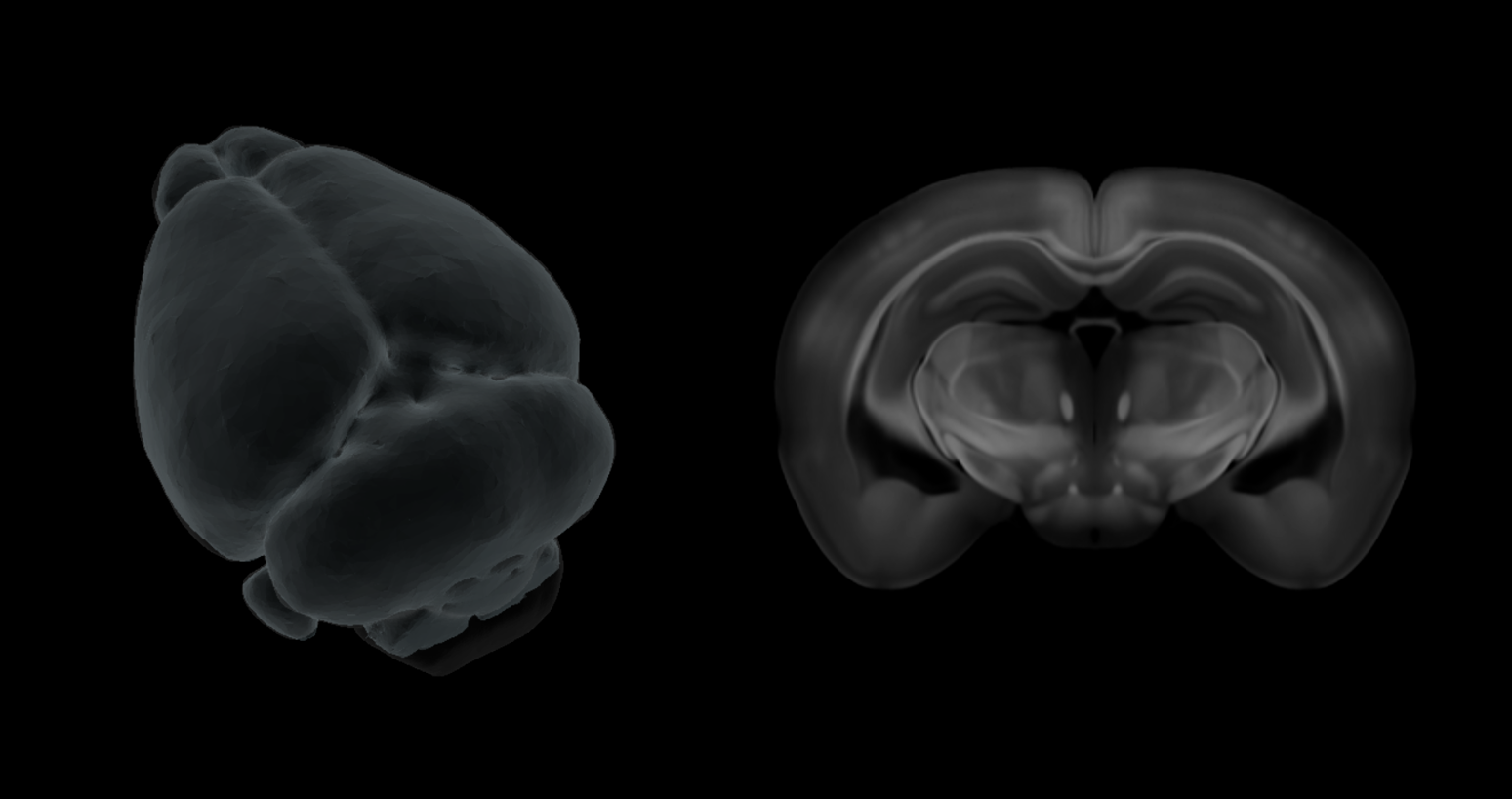
The Allen Mouse Brain Common Coordinate Framework (CCFv3) is built from serial two-photon tomography, a section-based imaging method. The Perens CCF was built differently: from intact, iDISCO+-cleared mouse brains imaged whole by light sheet microscopy, the same acquisition method LS-Journey™ uses to generate customer data.
Both are peer-reviewed, and both are legitimate references for their respective imaging methods. The practical question for any given dataset is which one was built the same way the data in hand was acquired.
Whole-brain c-Fos mapping counts activated cells per anatomical region across the whole brain, which means registration error is not spread evenly. It concentrates exactly at region boundaries, in curved or deep structures, and it compounds across hundreds of regions rather than the 10 to 15 a coarse region-of-interest analysis would track. An atlas built from a different imaging method shifts those boundaries first, and the cell counts follow.
This is also where the choice has the most practical weight, since a c-Fos study is usually run specifically to find which regions changed, not just that something changed somewhere. The semaglutide c-Fos map is one example of that kind of region-resolved result.
Four questions are enough to catch most mismatches before they reach the data:
Was it built from the same imaging method as your data? Section-based and light sheet atlases are not interchangeable references for the reasons above.
Does it cover adult mouse specifically, or is it adapted from another age or strain? A reference built for the animals you are actually imaging registers more predictably than one adapted from a different strain.
Is the region ontology detailed enough for your question? Ten to 15 broad regions are enough to say something changed in the midbrain, not which nucleus.
Is it peer-reviewed, and is the registration pipeline validated against it? A published atlas with an untested registration pipeline is only half the answer. Vibraint's image analysis module registers every LS-Journey™ dataset to the Perens CCF as one validated pipeline, not two separate steps.
Matching the atlas to the method solves registration. It does not, by itself, let you compare your data to anyone else's. That is where a shared coordinate framework becomes a spatial biology tool rather than just an imaging step: once your data and a public dataset are registered to the same reference brain, they overlay directly instead of each needing its own re-registration.
CNS-Voyager™ is built on that idea. It places LS-Journey™ results on the Perens CCF alongside Allen Brain connectivity data, spatial transcriptomics, and whole-genome Allen Brain Cell Atlas gene expression maps, plus stereotactic coordinate lookup, all on the same 3D framework. A brain-wide c-Fos map and a gene expression map only tell you something new together if they are standing on the same coordinates.
Not every use of a brain atlas needs this level of precision. A quick visual check of roughly where a structure sits tolerates a mismatched reference reasonably well. Quantitative, region-level analysis across many animals, the kind c-Fos studies and connectivity studies are built on, is exactly where atlas-method mismatch compounds instead of averaging out.
Whether that same-modality requirement holds as strictly for lower-resolution imaging methods such as MRI, as it does for whole-brain light sheet data remains unresolved, and worth testing directly by registering one dataset to both atlases side by side and comparing where the region boundaries actually land.
Match the atlas to the imaging method, not the other way around. A section-based atlas like the Allen CCF and a light sheet atlas like the Perens CCF are both valid references, but registration accuracy is highest when the atlas and the data were built the same way.
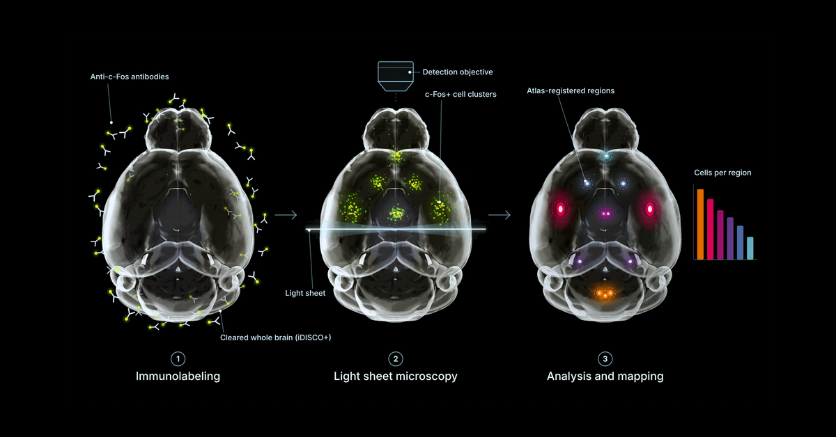
Vibraint's LS-Journey™ registers every dataset it generates (from immunolabeling through light sheet microscopy to AI-assisted analysis) to the peer-reviewed Perens CCF built specifically for that pipeline, then delivers the result on CNS-Voyager™ alongside Allen connectivity data, spatial transcriptomics, and whole-genome expression maps on the same coordinate framework.
A common coordinate framework, or CCF, is a 3D reference brain with every anatomical region defined and labeled, so that imaging data from different animals and different studies can be registered to the same coordinate system and directly compared.
Different imaging methods produce different tissue contrast, resolution, and deformation, particularly after tissue clearing, so an atlas built from one modality can register imperfectly to data acquired with another, shifting where region boundaries fall.
You can, but the Allen CCF was built from serial two-photon tomography, not light sheet microscopy of cleared tissue, so registration accuracy on cleared whole-brain light sheet data is better with an atlas built specifically for that method, such as the Perens CCF.
Yes. Whole-brain c-Fos mapping counts activated cells per anatomical region, so any registration error from an atlas-method mismatch is concentrated exactly where the activity signal is being measured, particularly near region boundaries.